Crystallography Sample Supports-MiTeGen晶体安装系统
上海金畔生物代理MiTeGen品牌蛋白结晶试剂耗材工具等,我们将竭诚为您服务,欢迎访问MiTeGen官网或者咨询我们获取更多相关MiTeGen品牌产品信息。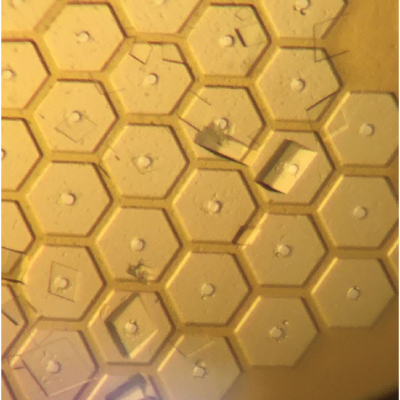

Crystallography Sample Supports
Crystallography Sample Supports will be a next-generation crystal mounting system that changes how crystals can be prepared for X-ray diffraction data collection.
This new system allows users to –
- Mount single crystals or thousands for micro-crystals
- Optimized for serial crystallography or single crystal XRD
- Cryogenic or Room Temperature diffraction
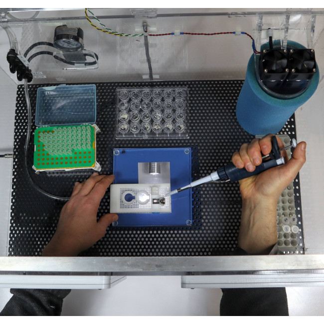
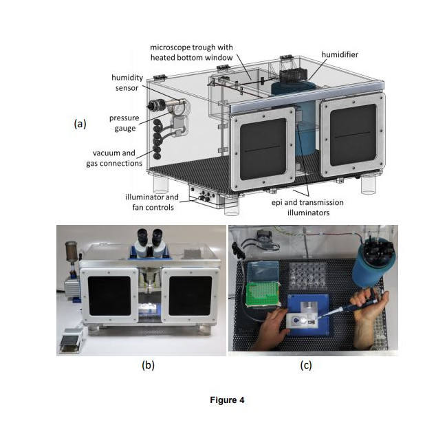
How it works – Crystals are mounted onto these supports utilizing a humidified sample loading box. The high-humidity box allows samples to be prepared without unintentional dehydration to the drops or crystals. The supports can than be plunge cooled (for cryo) or sealed (for room temperature work).
Primary system components –
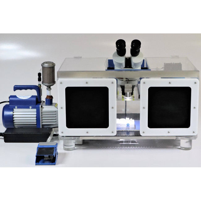
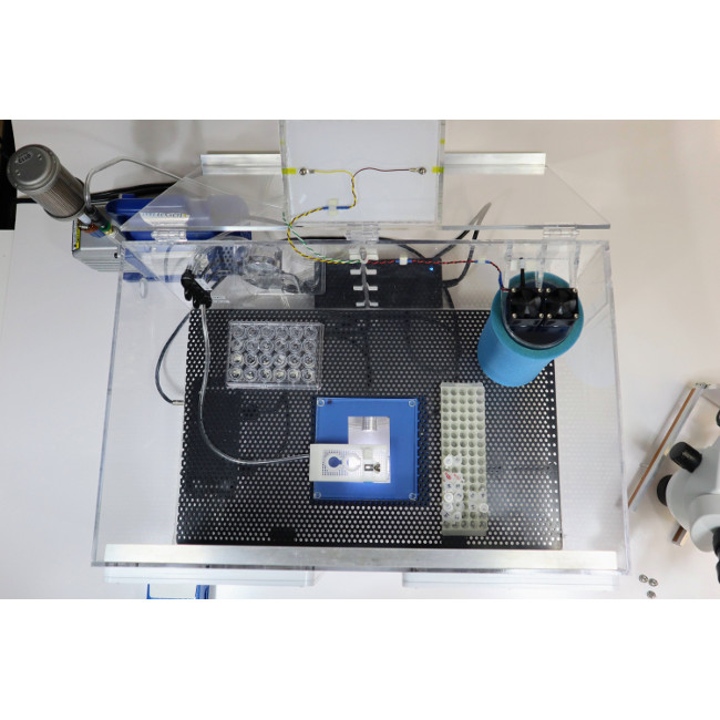
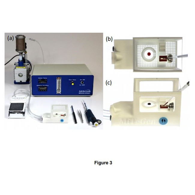
- Sample Loading Box – A humidified sample preparation box with optical microscope and vacuum sample wicking
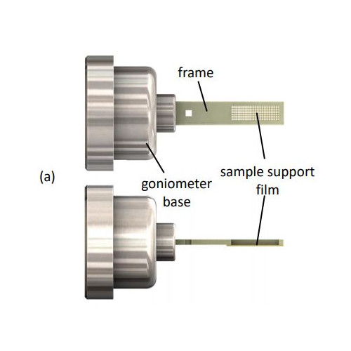
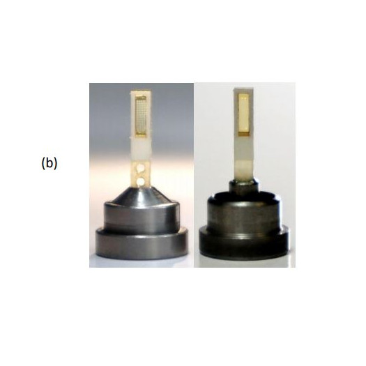
- Sample Supports – Sample Supports (Meshes) pre-mounted into automounter-compatible bases
- Sample Seals – Room temperature seals
- On-support crystallization plates (SSRL and Echo-compatible designs)
The system has multiple applications –
- Conventional Crystallography
- Serial Crystallography
- In Situ Crystallography
- Room Temperature Crystallography
- And more
晶体学样品支架
Crystallography Sample Supports 将是下一代晶体安装系统,它改变了晶体为 X 射线衍射数据收集而准备的方式。
这个新系统允许用户——
安装单晶或数千个微晶
针对串行晶体学或单晶 XRD 进行了优化
低温或室温衍射
工作原理 – 使用加湿的样品装载盒将晶体安装在这些支架上。高湿度盒允许制备样品而不会意外脱水成液滴或晶体。支撑物可以被骤冷(用于低温)或密封(用于室温工作)。
主要系统组件 –
样品装载盒 – 带有光学显微镜和真空样品芯吸功能的加湿样品制备盒
样品支架 – 样品支架(网格)预先安装到自动安装器兼容的底座中
样品密封 – 室温密封
支撑结晶板(SSRL 和 Echo 兼容设计)
该系统有多种应用——
常规晶体学
连续晶体学
原位晶体学
室温晶体学
和更多
Learn More About the Crystallography Sample Supports System
OR
Ask A Question
Product Information
- Key Features
- Learn About The Components
- Learn About The Use Cases
- Ask A Question
The Crystallography Sample Support system is an integrated system for preparing samples for several types of crystallography research. This system integrates and improves upon previously demonstrated concepts to deliver a versatile and high-performance solution.
类别:光束线和家庭源工具, 光束线样品制备, 低温晶体学, 晶体收获, 新产品, 室温筛选, 串行晶体学样品支持, 串行晶体学
产品信息
主要特点
了解组件
了解用例
问一个问题
晶体学样品支持系统是一个集成系统,用于为多种类型的晶体学研究制备样品。 该系统集成并改进了先前展示的概念,以提供多功能和高性能的解决方案。
The systems several salient features are:
- The sample supports are compatible with existing infrastructure for home source and mail-in SR crystallography, including sample handling tools, storage cassettes/pucks, automounters, and goniometer stages. This reduces overall cost and increases opportunities for data collection.
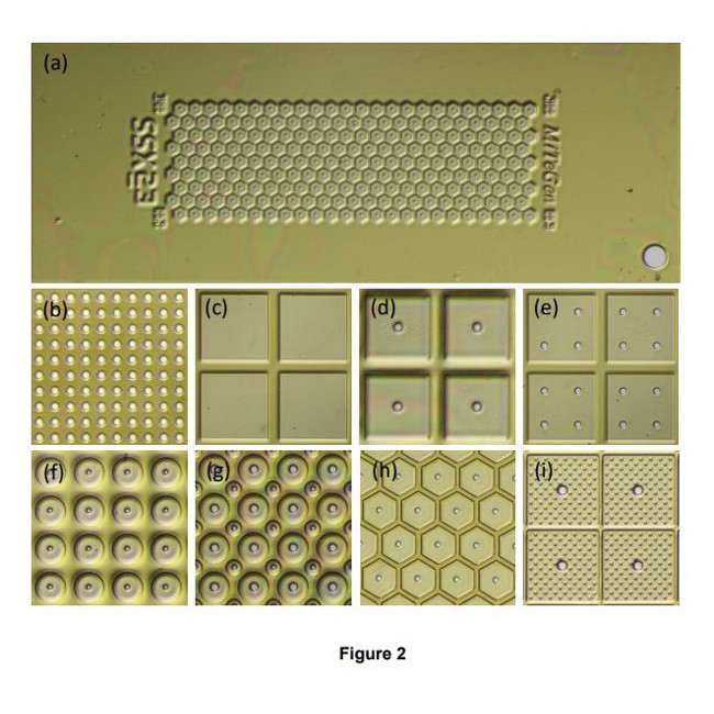
- A single sample holder platform accepts diverse sample support film designs for different size/shape crystals that aid in achieving regular or dispersed crystal positioning.
- Imaging of microcrystals as small as 1-2 µm over the entire active area of the support – not just in wells – is straightforward using both standard visible light microscopy and laser scanned nonlinear excitation modalities due the very thin polymer windows in each well, the thin polymer walls between wells, easy removal of crystal-contrast reducing surrounding liquid, and polyimide’s weak TPEF and zero SHG signals. IUCrJ BIOLOGY | MEDICINE research papers 23.
- With one or two stages of humidity control provided by the humidity-controlled sample loading station and the humidified glovebox, dehydration of crystallization drops and crystals – a critical issue when loading microcrystals for room temperature data collection – can be eliminated. As-grown crystal isomorphism is maintained, scaling and merging of diffraction data from many crystals is improved, and the number of crystals required for structure determination is reduced.
- Foot-pedal modulated suction within a humidified environment gives effective control of solution and crystal flows needed to achieve desired crystal positioning (e.g., random dispersion or at regularly spaced holes.) Positioning over holes can also be achieved by growing crystals in situ using vapor diffusion.
- Crystal position control is achieved using only thin polyimide films with micropatterned features only ~5-20 µm tall. Wells/walls defined by thick (130-250 m) substrates (e.g., silicon, polycarbonate) are not necessary. Background X-ray scatter is low at all positions on the sample support. Diffraction data can be collected from essentially all crystals on the support, not just those located over holes or windows, increasing efficiency of crystal use. X-rays can be incident from and diffracted through a wide angular range without encountering the support frame. Large sample support rotations eliminate data collection issues caused by preferential crystal orientation, and complete data sets can be collected from suitably large individual crystals.
- For room temperature data collection, ~4 µm Mylar sealing films provide adequate protection against dehydration for data collection on a timescale of ~1-2 hours. For room temperature storage and shipping, sealed samples can be stored with reservoir solution in vials, Eppendorf tubes, and multiwell plates.
- Crystal cryoprotection is simplified and made more effective. Cryoprotectantcontaining solutions (or oils) can be deposited on crystals on the sample support, and then withdrawn using suction through the support’s holes; repeating this deposition and removal two or more times can efficiently remove all solvent initially present on the crystal surface, eliminating the primary source of ice diffraction in cryocrystallography (Parkhurst et al., 2017; Moreau et al., 2020), and minimizing background scatter from excess cryoprotectant.
- For cryogenic temperature data collection, samples can be directly plunged in liquid nitrogen, and stored, shipped and handled in the same way and using the same tools as standard loops in cryocrystallography. IUCrJ BIOLOGY | MEDICINE research papers 24
- The supporting polyimide film is much thinner and has much less thermal mass per unit area than standard ~25 µm thick cryoEM grids. If ~1-2 µm crystals are used and excess solution carefully removed, cooling rates well in excess of 50,000 K/s (Kriminski et al., 2003; Warkentin et al., 2006), within a factor of ~10 of those achieved in typical cryoEM practice (>250,000 K/s), should be achievable using current state-of-the art LN2-based cooling methods. Consequently, capture via thermal quenching of roomtemperature biomolecular conformations should be nearly as effective using protein microcrystals as when using protein solutions on cryoEM grids (Kaledhonkar et al., 2019).
The Crystallography Sample Support system consists of several components, these are:
Sample Loading Box – A humidified sample preparation box with optical microscope and vacuum sample wicking
Sample Supports – Sample Supports (Meshes) pre-mounted into automounter-compatible bases
Sample Seals – Room temperature seals
On-support crystallization plates designs
The Crystallography Sample Support system has several use cases in crystallography. These include:
For conventional crystallography using nylon loops and microfabricated loops/mounts. Unlike for loop mounted crystals, there is no “fishing”: multiple crystals are simply deposited (with their mother liquor and any protein or PEG skins) on the support film. Excess liquid is easily removed, background scatter is reduced, and cooling rates are increased. Crystals are confined to a plane adjacent to the support film, simplifying imaging and reducing the chance that the X-ray beam illuminates more than one crystal. Cryoprotection is conveniently performed on the support, with little risk of crystal damage or loss.
For in situ crystallography, the present sample supports have an advantage over some goniometer-base-mounted crystallization devices of being fully compatible with beamline automounters and other high-throughput hardware. The present system also allows mother liquor to be suctioned off after crystallization without risk of crystal damage or dehydration to reduce background scatter and simplify cryocooling.
For serial synchrotron crystallography the Crystallography Sample Support provides a comprehensive and highly flexible solution. Moreover, because sample preparation and handling is simplified, sample damage and dehydration are minimized, sample imaging is improved, background scatter is reduced, cryoprotection is simplified, and cooling rates are maximized, this system has excellent potential to replace conventional loops and mounts and associated protocols in a large fraction of biomolecular crystallography applications. Structure determinations, especially at room temperature where crystallography maintains a major advantage relative to cryoEM — typically require not just one crystal but many, and that the crystals be highly isomorphous. The present integrated system is optimized for this task and will allow much more efficient use of crystals and synchrotron beamtime.
For room temperature crystallography the one or two stages of humidity control provided by the humidity-controlled sample loading station and the humidified glovebox, dehydration of crystallization drops and crystals – a critical issue when loading microcrystals for room temperature data collection – can be eliminated. As-grown crystal isomorphism is maintained, scaling and merging of diffraction data from many crystals is improved, and the number of crystals required for structure determination is reduced. In addition for room temperature data collection, ~4 µm Mylar sealing films provide adequate protection against dehydration for data collection on a timescale of ~1-2 hours. For room temperature storage and shipping, sealed samples can be stored with reservoir solution in vials, Eppendorf tubes, and multiwell plates.
Do you have a question concerning how the Crystallography Sample Support system might apply to your research? Contact us using the contact form found under the far right hand side tab.
Want to learn more about the Crystallography Sample Support system? Contact us using the form below and we can collaborate on how the system can be used to improve your research.
该系统的几个显着特点是:
样品支架与用于家庭源和邮寄 SR 晶体学的现有基础设施兼容,包括样品处理工具、存储盒/圆盘、自动安装器和测角仪阶段。这降低了总体成本并增加了数据收集的机会。
单个样品架平台接受不同尺寸/形状晶体的不同样品支撑膜设计,有助于实现规则或分散的晶体定位。
由于每个孔中的聚合物窗口非常薄,不仅在孔中,而且使用标准可见光显微镜和激光扫描非线性激发模式,在整个载体活性区域上对小至 1-2 µm 的微晶进行成像都很简单。孔之间的聚合物壁,易于去除降低晶体对比度的周围液体,以及聚酰亚胺的弱 TPEF 和零 SHG 信号。 IUCrJ 生物学 |医学研究论文 23.
通过湿度控制的样品加载站和加湿手套箱提供的一到两个阶段的湿度控制,可以消除结晶液滴和晶体的脱水——加载微晶进行室温数据收集时的一个关键问题——可以消除。随着生长的晶体同构性得以保持,来自许多晶体的衍射数据的缩放和合并得到改进,并且结构测定所需的晶体数量减少。
加湿环境中的脚踏式调制吸力可有效控制溶液和晶体流动,以实现所需的晶体定位(例如,随机分散或在规则间隔的孔中)。也可以通过使用蒸汽扩散原位生长晶体来实现孔上的定位.
仅使用具有仅 ~5-20 µm 高的微图案特征的薄聚酰亚胺薄膜即可实现晶体位置控制。不需要由厚 (130-250 m) 基材(例如硅、聚碳酸酯)定义的孔/壁。样品支架上所有位置的背景 X 射线散射都很低。衍射数据可以从载体上的基本上所有晶体中收集,而不仅仅是那些位于孔或窗上的晶体,从而提高了晶体的使用效率。 X 射线可以从很宽的角度范围入射和衍射,而不会遇到支撑框架。大样品支架旋转消除了由优先晶体取向引起的数据收集问题,并且可以从适当大的单个晶体中收集完整的数据集。
对于室温数据收集,~4 µm 聚酯薄膜密封膜可在~1-2 小时的时间范围内为数据收集提供足够的脱水保护。对于室温储存和运输,密封样品可以与储存液一起储存在小瓶、Eppendorf 管和多孔板中。
晶体冷冻保护被简化并变得更有效。含有冷冻保护剂的溶液(或油)可以沉积在样品支架上的晶体上,然后通过支架的孔抽吸;重复这种沉积和去除两次或更多次可以有效地去除最初存在于晶体表面的所有溶剂,消除低温晶体学中冰衍射的主要来源(Parkhurst 等人,2017 年;Moreau 等人,2020 年),并最大限度地减少背景散射来自过量的冷冻保护剂。
对于低温温度数据收集,样品可以直接浸入液氮中,并以与低温晶体学中的标准回路相同的方式和工具进行储存、运输和处理。 IUCrJ 生物学 |医学研究论文 24
与标准 ~25 µm 厚的冷冻电镜网格相比,支撑聚酰亚胺薄膜要薄得多,单位面积的热质量要少得多。如果使用 ~1-2 µm 晶体并小心去除多余的溶液,冷却速率将远远超过 50,000 K/s(Kriminski 等人,2003 年;Warkentin 等人,2006 年),在所达到的约 10 倍以内在典型的冷冻电镜实践 (>250,000 K/s) 中,使用当前最先进的基于 LN2 的冷却方法应该可以实现。因此,使用蛋白质微晶通过室温生物分子构象的热猝灭进行捕获应该几乎与在冷冻电镜网格上使用蛋白质溶液一样有效(Kaledhonkar 等人,2019 年)。
晶体学样品支持系统由几个组件组成,它们是:
样品装载盒 – 带有光学显微镜和真空样品芯吸功能的加湿样品制备盒
样品支架 – 样品支架(网格)预先安装到自动安装器兼容的底座中
样品密封 – 室温密封
支撑结晶板设计
晶体学样品支持系统在晶体学中有几个用例。这些包括:
对于使用尼龙环和微加工环/支架的传统晶体学。与循环安装的晶体不同,没有“钓鱼”:简单地将多个晶体(连同它们的母液和任何蛋白质或 PEG 皮)沉积在 sup 上
